
Pictures of Cysts: Types, Symptoms & What to Look For
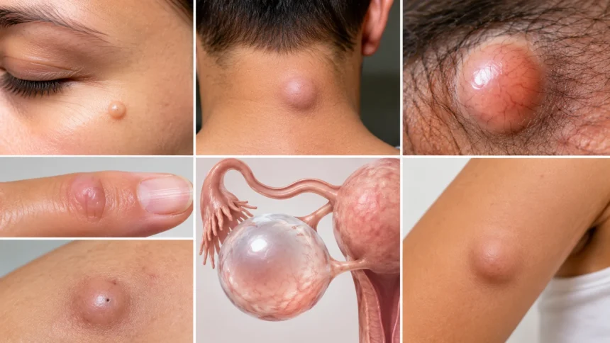
Cysts are closed or partly enclosed sacs that can develop in the skin, under the skin, or inside organs and tissues. Depending on the type, a cyst may contain fluid, keratin, pus, blood, air, or another material, which means two cysts can look and feel very different. When people search for pictures of cysts, they are often trying to decide whether a new bump, swelling, or lump resembles something harmless or something that needs medical attention. Photos can provide useful visual context, but they cannot reliably diagnose a lump because cysts can resemble boils, lipomas, swollen lymph nodes, abscesses, tumors, and other skin or soft-tissue conditions. Size, location, tenderness, growth pattern, skin changes, and associated symptoms all matter. A healthcare professional may need to examine the area directly or use imaging to determine what it actually is.
Some cysts remain small and painless for years, while others become inflamed, infected, uncomfortable, or large enough to interfere with movement. Epidermoid and pilar cysts commonly develop beneath the skin, ganglion cysts often appear near joints, and pilonidal cysts occur near the crease of the buttocks. Other cysts develop deeper inside the body, such as ovarian or breast cysts, where appearance on the skin may reveal little or nothing. The most useful way to interpret cyst pictures is therefore to understand typical patterns rather than searching for an exact visual match. This guide explains common types of cysts, how they may look and feel, symptoms that can accompany them, and warning signs that deserve medical evaluation. It also explains why squeezing or draining an unidentified cyst at home can sometimes create more problems than it solves.
What Does a Cyst Look Like?
A skin cyst often looks like a rounded or dome-shaped bump beneath the surface of the skin. It may be close to the normal skin color, slightly yellow, pink, red, or darker depending on inflammation and the person’s natural skin tone. Some cysts feel smooth and movable when gently touched, while others seem more firmly attached to surrounding tissue. The skin over the lump may remain completely normal when the cyst is not irritated. In photographs, uncomplicated cysts may therefore look surprisingly subtle, especially when they are small. A visible bump alone cannot confirm that a lesion is a cyst because many other conditions produce similar shapes. Location, texture, symptoms, duration, and changes over time are equally important when evaluating a lump.
Many epidermoid cysts have a small dark opening or central punctum on the surface, although this feature is not always visible. The opening represents a connection with the skin surface and may resemble a clogged pore. If the cyst becomes damaged or squeezed, thick whitish or yellow material with an unpleasant odor may come out. This material is usually keratin rather than ordinary pus when the cyst is not infected. A picture showing a central opening can suggest an epidermoid cyst, but it should not be treated as definitive proof. Other blocked follicles and enlarged pores can appear similar. Repeated squeezing can inflame the cyst wall and make the area larger, redder, and more difficult to treat.
Inflamed cysts often look very different from quiet cysts. The skin can become red or deeply discolored, swollen, shiny, warm, and tender, and the lump may increase in size over a short period. In darker skin tones, inflammation may appear purple, gray, brown, or deeper than the surrounding skin rather than bright red. An inflamed cyst can resemble an abscess, boil, or infected hair follicle, especially when pain becomes significant. Some inflamed cysts are sterile, meaning inflammation is present without a bacterial infection. Others can become secondarily infected and produce drainage or worsening tenderness. Because appearance alone cannot reliably distinguish these situations, a rapidly painful or enlarging lump may need professional assessment rather than home treatment based on photographs.
A cyst can feel soft, firm, rubbery, or fluctuant depending on what it contains and where it is located. A fluid-filled cyst may feel as though there is movement beneath the fingers, while a keratin-filled cyst can feel denser. Ganglion cysts near the wrist may feel firm despite containing a jelly-like fluid. Deep cysts may not be clearly visible at all and may be detected only as a lump or through imaging. The consistency of a lump can provide useful diagnostic clues, but self-examination has limits. People often have difficulty deciding whether a lesion is truly movable or attached. Any lump that is hard, fixed, persistently enlarging, or associated with unexplained systemic symptoms deserves medical evaluation rather than being assumed to be a simple cyst.
Size also varies significantly. A cyst can be smaller than a pea or grow several centimeters across, and some internal cysts become much larger before producing symptoms. Size alone does not determine whether a cyst is dangerous. A tiny cyst near a sensitive structure can cause significant discomfort, while a larger superficial cyst may remain painless. However, rapid enlargement is more concerning than a lump that has remained unchanged for years. Photographs taken weeks apart can sometimes help document change more accurately than memory alone. If you are monitoring a visible lump, use consistent lighting and include a size reference without repeatedly pressing or manipulating the area. Changes in shape, color, drainage, pain, or surrounding skin are often more informative than a single picture.
Epidermoid and “Sebaceous” Cysts
Epidermoid cysts are among the most common skin cysts and frequently appear in online pictures of cysts. They usually develop when skin cells that would normally shed become trapped deeper in the skin and continue producing keratin. The resulting lump is commonly round, slow-growing, and located just beneath the skin. Typical areas include the face, neck, chest, back, trunk, and sometimes the genital region. The surface may be normal in color, and a central punctum may be visible. Many people refer to these lesions as sebaceous cysts, although most common cysts described that way are actually epidermoid cysts rather than true cysts arising from sebaceous glands. The distinction matters mainly for accurate medical terminology rather than everyday symptom recognition.
An uncomplicated epidermoid cyst is usually painless. It may feel smooth and somewhat mobile beneath the skin and can remain nearly unchanged for months or years. Some people notice the cyst only because it becomes visible in certain lighting or when clothing rubs against it. Larger cysts can become uncomfortable because of their location, particularly on the back, neck, or areas exposed to pressure. The contents are often thick and paste-like because the sac contains keratin from trapped skin cells. If material leaks through the surface opening, it may have a strong odor. This does not automatically mean the cyst is infected. However, spontaneous rupture beneath the skin can produce intense inflammation even without bacteria.
When an epidermoid cyst becomes inflamed, it can rapidly become tender, swollen, and discolored. People sometimes assume this change means the cyst has suddenly become dangerous, but inflammation can occur after friction, minor trauma, or rupture of the cyst wall. The immune system reacts strongly when keratin escapes into surrounding tissue. This reaction can make the lesion look much larger than it did previously. A clinician may sometimes treat inflammation first and delay complete removal until the area has settled. Attempting to squeeze an inflamed cyst can force material deeper into the tissue and increase irritation. It may also damage the cyst wall, making later removal more difficult. Gentle care and medical evaluation are generally safer when pain or swelling is significant.
Treatment is not always required for a small, painless epidermoid cyst. If the diagnosis is clear and the cyst is not causing discomfort, some people choose simple observation. Removal may be considered when the lump is recurrently inflamed, painful, enlarging, cosmetically bothersome, or located where friction repeatedly irritates it. Complete surgical removal aims to remove both the contents and the cyst wall because leaving part of the capsule behind can allow recurrence. Simply draining the contents may temporarily flatten the cyst without permanently eliminating it. The exact procedure depends on location, size, and inflammation. People should avoid cutting into a cyst at home because this can cause infection, bleeding, scarring, and incomplete drainage.
Any presumed epidermoid cyst that behaves unusually should be reassessed. Warning signs include persistent rapid growth, marked firmness, fixation to deeper tissues, repeated bleeding, an unusual ulcerated surface, or a diagnosis that remains uncertain. Most epidermoid cysts are benign, but assuming every lump with a round shape is harmless can delay diagnosis of other conditions. A dermatologist or other clinician can often recognize typical cysts through examination. Ultrasound or tissue analysis may occasionally be used when the diagnosis is unclear. If a removed cyst has an unusual appearance, it may be sent for laboratory examination. The important point is not to become alarmed by every skin lump but to recognize when a familiar-looking cyst no longer follows a typical pattern.
Pilar Cysts on the Scalp
Pilar cysts, also called trichilemmal cysts, commonly develop on the scalp and arise from the outer root sheath of hair follicles. They often appear as smooth, firm, rounded lumps beneath normal-looking scalp skin. Unlike many epidermoid cysts, they usually do not have a visible central punctum. A person may discover one while washing, brushing, or styling their hair rather than seeing it directly in a mirror. Because the scalp contains hair and curved surfaces, pilar cysts can be difficult to photograph clearly. They may appear as a raised area under the hair rather than an obvious skin lesion. Some people develop more than one, and a tendency toward pilar cysts can run in families.
Most pilar cysts grow slowly and cause no pain. They may feel firmer than people expect because the keratin inside is often densely packed. The cyst can move slightly beneath the skin, although larger ones may feel less mobile. Hairstyles, helmets, hats, or combing can make a scalp cyst noticeable if pressure is applied repeatedly. A cyst located where the head rests on a pillow or chair may also become irritating over time. The overlying skin usually remains unchanged unless the cyst becomes inflamed or traumatized. Because scalp lumps can also represent lipomas, inflamed follicles, bony prominences, and other conditions, a physical examination is useful when the diagnosis is uncertain.
Pilar cysts can occasionally rupture and become inflamed. When this happens, the area may become tender, swollen, warm, and discolored, creating an appearance similar to an abscess. Trauma from repeated picking or attempting to puncture the cyst can increase this risk. Keratin may escape into surrounding tissue and trigger a substantial inflammatory reaction. Medical management depends on whether there is simple inflammation, infection, or another complication. People should not repeatedly press a painful scalp lump because this can worsen discomfort and swelling. If fever, spreading redness, drainage, or significant pain develops, prompt evaluation is sensible. The clinician can determine whether treatment beyond observation is needed.
Removal can be considered for pilar cysts that are bothersome, recurrently inflamed, enlarging, or cosmetically concerning. Because these cysts often have a relatively well-defined wall, clinicians can frequently remove them completely through a small surgical procedure. Local anesthesia is commonly used for superficial lesions. The scalp has a rich blood supply, so careful technique is important even for minor procedures. Hair usually does not need extensive removal around a small cyst, although specific preparation varies. Once the capsule is removed, recurrence at that exact site is less likely, although someone predisposed to pilar cysts may develop new cysts elsewhere. Follow-up instructions usually focus on wound care, bleeding, and signs of infection.
Very rapidly growing scalp cysts deserve attention rather than being assumed to be ordinary pilar cysts. Rare proliferating forms can become much larger and behave differently from typical small lesions. A sudden increase in size, ulceration, repeated bleeding, or significant change in a long-standing lump warrants professional evaluation. Most scalp cysts remain benign, so these warning signs should be understood as reasons to check rather than reasons to panic. A dermatologist, surgeon, or primary care clinician can examine the lesion and decide whether removal or further testing is appropriate. Photographs may document growth, but they cannot evaluate depth or tissue characteristics. Any scalp lump that is changing significantly should therefore be assessed directly rather than managed solely through visual comparison online.
Ganglion Cysts on the Wrist, Hand, or Foot
Ganglion cysts commonly develop near joints or tendons, especially around the wrist and hand. They contain a thick, jelly-like fluid associated with joint or tendon structures rather than keratin from skin cells. In pictures, a ganglion cyst often appears as a smooth, rounded swelling that becomes more visible when the wrist is bent. The overlying skin usually looks normal because the cyst sits beneath it. Some ganglion cysts are very small and can be felt more easily than seen. Others become prominent enough to change the contour of the wrist or hand. Their size may fluctuate depending on joint activity, and a cyst can occasionally become smaller or disappear without treatment.
Symptoms range from none at all to aching, pressure, weakness, or discomfort with certain movements. A ganglion near a nerve can cause tingling, numbness, or pain if it compresses nearby structures. People who repeatedly use their hands for sports, typing, tools, or certain occupations may notice the lump more when activity increases. However, everyday use does not necessarily cause the cyst. Diagnosis is often based on location and examination, although ultrasound or other imaging can be used when the lump is unusual. Some ganglion cysts feel quite firm, which can make people worry that the lump is solid. Their firmness does not automatically indicate a dangerous growth because the fluid inside can be thick and the surrounding capsule tense.
One traditional home remedy involved hitting a wrist ganglion with a heavy book to make it rupture. This approach is unsafe and should not be used because it can injure bones, nerves, tendons, blood vessels, and surrounding soft tissue. Puncturing the cyst at home is also risky because infection can be introduced into tissues near a joint. If the ganglion is painless and does not interfere with function, observation may be appropriate. Some cysts shrink without intervention. Activity modification or a temporary brace may help with discomfort in selected cases. The best approach depends on symptoms and location. A medical professional can confirm that the swelling actually resembles a ganglion before treatment decisions are made.
Aspiration is one treatment option in which a clinician uses a sterile needle to remove fluid from the cyst. This can reduce its size and relieve pressure, but recurrence is relatively common because the connection with the joint or tendon may remain. Some ganglion cysts are more difficult to aspirate because their contents are thick or their location makes needle treatment less appropriate. Surgical removal may be considered when the cyst repeatedly returns, causes significant pain, compresses nerves, or limits movement. Surgery aims to remove the cyst and its connection to the surrounding tissue. Even after surgery, recurrence can occur. Treatment therefore focuses mainly on symptoms and function rather than removing every ganglion cyst simply because it is visible.
Not every lump near the wrist is a ganglion cyst. Bone growths, tendon swelling, lipomas, vascular abnormalities, arthritis-related changes, and other masses can appear in similar locations. A lump that is pulsating, unusually hard, rapidly enlarging, associated with significant weakness, or accompanied by severe pain should be assessed rather than punctured or manipulated. Injury-related swelling also needs appropriate evaluation because a fracture or tendon problem can sometimes create a visible bump. Photographs of ganglion cysts can help people recognize the typical smooth contour, but touch, movement, and location relative to the joint provide important clues that pictures cannot show. When hand function or nerve symptoms are affected, timely professional evaluation is particularly important.
Pilonidal Cysts Near the Tailbone
A pilonidal cyst develops in the cleft between the buttocks, usually near the tailbone. It often begins when hair, friction, and skin debris contribute to inflammation beneath the skin in that area. Some people have small pits or openings in the skin without significant symptoms, while others develop painful abscesses. Because of the sensitive location, people may search for pictures before deciding whether the problem needs medical attention. A pilonidal cyst may appear as a small dimple, swollen lump, draining opening, or inflamed area near the top of the buttock crease. Visual appearance varies considerably depending on whether the condition is quiet, chronically draining, or acutely infected.
When a pilonidal cyst becomes infected or forms an abscess, pain can develop quickly. Sitting may become difficult, and the area can become swollen, warm, red or darkened, and extremely tender. Pus or blood may drain from a small opening, sometimes with an unpleasant odor. Fever is possible but not always present. Because the cyst is located in a warm area exposed to pressure and friction, inflammation can become quite uncomfortable. People sometimes mistake the early symptoms for a pimple or ingrown hair. However, a rapidly enlarging painful lump in the buttock crease is more consistent with a pilonidal abscess and usually needs clinical evaluation rather than repeated squeezing or application of random topical products.
Acute pilonidal abscesses often require drainage by a healthcare professional. Antibiotics alone may not be enough when a significant pocket of pus has formed because the infected material needs to escape. The exact technique depends on the size and severity of the abscess. After drainage, wound care and follow-up are important because pilonidal disease can recur. Some people experience only one episode, while others develop chronic draining sinuses or repeated infections. Persistent disease may require a surgical procedure designed to remove affected tissue or reshape the area to reduce recurrence. Several surgical approaches exist, so treatment should be individualized based on anatomy, disease extent, prior procedures, and specialist experience.
Hair management and reducing prolonged moisture or friction may be recommended as part of recurrence prevention. Depending on the individual, clinicians may discuss careful trimming, other hair-removal methods, hygiene, and avoiding prolonged pressure. Recommendations can differ, particularly after surgery, so patients should follow the instructions given for their specific treatment. Weight, body shape, occupation, and sitting time can affect the local environment but are not reasons for blame. Pilonidal disease can affect otherwise healthy people and is especially common in younger adults. Wearing comfortable clothing and keeping the area dry may help reduce irritation. However, preventive measures cannot guarantee that another episode will not occur, especially when deeper sinus tracts have already developed.
A pilonidal cyst is usually recognizable by its location, but other conditions can affect the same area. Hidradenitis suppurativa, skin abscesses, infected follicles, and other inflammatory disorders can produce painful lumps or drainage around the buttocks. A clinician may need to examine the exact location and pattern before confirming the diagnosis. Severe pain, spreading redness, fever, significant drainage, or rapidly increasing swelling warrants prompt medical attention. Recurrent lesions in multiple areas such as the groin, armpits, or buttocks may suggest another inflammatory skin condition. Photographs can help document progression, but intimate images should be stored and shared carefully to protect privacy. Direct examination remains the safest way to distinguish recurrent pilonidal disease from similar-looking conditions.
Cystic Acne and Acne Cysts
Cystic acne refers to severe inflammatory acne that produces deep, painful lumps beneath the skin. These lesions are not identical to a classic epidermoid cyst, but they are commonly called acne cysts because of their deep, swollen appearance. They often occur on the face, jawline, chest, shoulders, or back and may remain painful for days or weeks. In pictures, cystic acne can appear as large red, purple, brown, or deeply discolored bumps without a visible whitehead. The inflammation develops deeper in the skin than an ordinary pimple, which makes squeezing especially ineffective. Pressing the lesion can drive inflammation further into surrounding tissue and increase the risk of scarring or post-inflammatory discoloration.
Deep acne nodules and cyst-like lesions can feel firm or fluctuant, and several may occur together. The surrounding skin may also contain blackheads, whiteheads, smaller inflamed pimples, and older scars. People with darker skin tones may notice persistent brown or gray marks long after active inflammation settles. Because severe acne affects appearance and can be painful, it may have a meaningful emotional impact as well. Treatment should therefore consider both physical and psychological wellbeing. Over-the-counter products may help mild acne but can be insufficient for frequent deep lesions. A dermatologist can determine whether prescription topical medication, oral therapy, hormonal treatment, or another approach is appropriate based on severity and individual health factors.
Picking or puncturing cystic acne increases the likelihood of scarring. Unlike a superficial whitehead, a deep acne lesion may have no clear pathway to the skin surface. Applying pressure can rupture the inflamed follicle internally and spread inflammatory material into nearby tissue. This can make the bump appear temporarily larger and more painful. Gentle skin care is generally more useful than aggressive scrubbing because harsh products can irritate the skin barrier without reaching the deep inflammation. Non-comedogenic moisturizers and sunscreen may also be important, especially when acne treatments cause dryness or increase sensitivity. People often use multiple strong products at once in an attempt to speed improvement, but excessive irritation can make the overall complexion more difficult to manage.
Certain large acne lesions can sometimes be treated directly by a dermatologist when rapid improvement is important. For example, a clinician may inject medication into a particularly painful inflammatory lesion to reduce swelling. This is different from attempting to drain it at home. Long-term control usually requires treating the acne process that causes repeated lesions rather than dealing with each cyst individually. Consistency matters because many acne medications take weeks to produce meaningful improvement. Hormonal patterns, medications, skin-care products, and other factors can influence acne severity. Scarring risk is one reason not to wait indefinitely when deep painful lesions are occurring frequently. Early effective treatment may reduce both active breakouts and permanent changes in skin texture.
A sudden onset of severe acne-like cysts can occasionally have another explanation, particularly when accompanied by unusual symptoms or medication changes. Folliculitis, hidradenitis suppurativa, rosacea variants, infections, and other inflammatory skin disorders can sometimes resemble acne. Lesions concentrated in unusual areas or recurring as tunnels and draining wounds deserve dermatologic evaluation. Pictures can help demonstrate how outbreaks change over time, but they should not replace examination when the diagnosis remains uncertain. People should also avoid judging acne severity solely by redness because inflammation can appear differently across skin tones. Pain, depth, number of lesions, scarring, and recurrence all matter. Accurate diagnosis allows treatment to target the underlying process rather than simply treating every deep bump as a generic cyst.
Breast, Ovarian, and Other Internal Cysts
Breast cysts are fluid-filled sacs within breast tissue and are particularly common before menopause, although they can occur at other ages. Unlike skin cysts, they may not create a visible surface bump unless they are relatively large or close to the skin. A breast cyst can feel smooth, round, or oval and may be tender, especially around hormonal changes. Some fluctuate in size during the menstrual cycle. Because many different breast conditions can cause lumps, self-examination cannot reliably determine whether a new lump is a cyst. Clinical breast examination and imaging such as ultrasound or mammography may be used based on age and circumstances. Any new or persistent breast lump deserves appropriate assessment rather than being diagnosed from pictures or touch alone.
Simple breast cysts seen on imaging are generally fluid-filled and may not require treatment if they are small and painless. A clinician can sometimes drain a bothersome cyst using a needle, which may also help confirm its fluid-filled nature. If fluid is bloody, the lump does not disappear, or imaging has unusual features, further evaluation may be necessary. Complex or complicated cystic findings are managed according to their specific imaging characteristics rather than simply being labeled harmless. People should also seek assessment for nipple changes, skin dimpling, unexplained breast redness, persistent focal pain, or a lump that continues to enlarge. Most breast lumps are not cancer, but visual appearance alone cannot distinguish benign and malignant causes reliably.
Ovarian cysts develop on or within an ovary and cannot usually be seen externally. Many form as part of the normal menstrual cycle and resolve without treatment. Others arise from different tissues or medical conditions and may persist. Symptoms can include pelvic pressure, one-sided lower abdominal pain, bloating, discomfort during sex, or changes in menstrual symptoms, although many ovarian cysts cause no symptoms. Ultrasound is commonly used to examine their size and internal appearance. People searching for pictures of ovarian cysts are often viewing medical imaging rather than ordinary photographs. Interpreting those images requires training because features such as internal walls, solid areas, blood flow, and fluid characteristics influence how clinicians assess a cyst.
Most ovarian cysts do not represent emergencies, but sudden severe pelvic or abdominal pain deserves prompt evaluation. A cyst can occasionally rupture or contribute to ovarian torsion, in which the ovary twists and its blood supply becomes compromised. Severe one-sided pain accompanied by nausea or vomiting is particularly concerning for torsion. Dizziness, fainting, significant weakness, or signs of internal bleeding after sudden pain also require urgent care. These complications cannot be diagnosed by looking at the abdomen or comparing symptoms with internet photographs. Pregnancy status can also affect evaluation of pelvic pain. Anyone with severe or rapidly worsening symptoms should seek urgent medical assessment rather than waiting for a routine appointment.
Cysts can develop in many other organs, including the kidneys, liver, pancreas, and thyroid. Some are discovered incidentally during imaging performed for unrelated reasons and never cause symptoms. Others require follow-up because of their size, internal characteristics, growth, or effect on nearby structures. The meaning of the word “cyst” therefore depends heavily on the organ involved. A simple kidney cyst in an older adult is very different from an inflamed skin cyst or a complex pancreatic cyst. Internet photographs provide little useful information for internal findings because interpretation depends on ultrasound, CT, MRI, laboratory results, and clinical context. If imaging has identified an internal cyst, the most useful questions are what type it appears to be, whether follow-up is needed, and which symptoms should prompt earlier reassessment.
Signs a Cyst May Be Infected or Inflamed
Increasing pain is one of the clearest signs that a previously quiet cyst has changed. A lump that has been painless for months and suddenly becomes tender may have ruptured internally, become inflamed, or developed an infection. Pain can range from mild soreness to throbbing discomfort that makes clothing or movement difficult. The surrounding skin may also become warmer than nearby tissue. These changes are particularly common with epidermoid and pilonidal cysts. Not every inflamed cyst contains bacteria, so antibiotics are not automatically required in every situation. A clinician evaluates the appearance, drainage, symptoms, and examination findings to determine whether infection is likely. Rapidly worsening pain should not be managed by repeatedly squeezing the lump in an attempt to relieve pressure.
Redness or other color change can accompany inflammation, although appearance varies considerably with skin tone. On lighter skin, the area may appear pink or bright red. On darker skin, inflammation may be seen as purple, deep brown, gray, or a more subtle change compared with surrounding skin. Swelling and shiny stretched skin can provide additional clues when color changes are difficult to appreciate. Spreading discoloration around a painful lump may indicate inflammation extending beyond the cyst. Marking the edge of redness is sometimes used clinically to monitor progression, but home monitoring should not delay care when symptoms are rapidly worsening. A cyst associated with expanding warmth, tenderness, or systemic illness requires more attention than one that remains stable and painless.
Drainage may occur when a cyst opens spontaneously or after pressure damages the surface. Thick, whitish, cheesy material is typical of keratin-filled cysts and may have a strong smell. Pus is more suggestive of infection, although visual appearance cannot always distinguish the two. Bloody drainage can occur when inflamed tissue is fragile or has been repeatedly manipulated. Any drainage should be handled with clean dressings, and hands should be washed before and after touching the area. Sharing towels or repeatedly applying nonsterile tools increases infection risk. If drainage continues, the wound becomes larger, or pain remains substantial, medical evaluation may be needed. A cyst that empties through the surface can recur because the capsule often remains beneath the skin.
Fever, chills, worsening fatigue, or feeling generally unwell raises concern that an infection may be extending beyond a localized skin problem. These symptoms are not typical of a small uncomplicated cyst. People with diabetes, immune suppression, poor circulation, or certain chronic illnesses may be at greater risk of complications from skin infections and should have a lower threshold for seeking care. Location also matters. Infections around the face, spine, genitals, hands, or near important joints may require particularly careful assessment. A clinician may need to drain an abscess, prescribe medication, or obtain a sample when infection is suspected. Trying to manage a severe infection entirely with home remedies can allow it to progress.
Repeated inflammation deserves evaluation even if each episode settles eventually. A cyst that swells every few months may have an intact wall that continues refilling, or the diagnosis may be something other than a simple cyst. Hidradenitis suppurativa, recurrent folliculitis, and other conditions can cause repeated painful lumps that are sometimes mistaken for cysts. Persistent drainage, tunnels under the skin, or lesions occurring repeatedly in the armpits, groin, or beneath the breasts are particularly important clues. Removing an individual cyst may solve one localized problem, but recurring lesions in multiple locations may require a broader treatment plan. Keeping a brief record of when lesions appear and where they occur can help clinicians identify patterns that are difficult to remember during a single appointment.
When Should You Worry About a Cyst?
Rapid growth is one reason to have a presumed cyst evaluated. Many benign cysts enlarge gradually, sometimes over years, so a lump that noticeably changes over days or weeks deserves more attention. Rapid growth does not automatically mean cancer because inflammation, infection, or bleeding can also increase size quickly. However, the cause should be clarified rather than assumed. Measuring the lump occasionally or taking dated photographs can help demonstrate the rate of change. Avoid checking it dozens of times per day because repeated pressing can irritate the tissue. A clinician can determine whether the lesion has features typical of a cyst or whether ultrasound, imaging, removal, or tissue sampling would be useful.
A hard or fixed lump should also be assessed, particularly when it does not move relative to deeper tissues. Some normal cysts feel firm, so firmness alone is not diagnostic. The concern increases when the lump has an irregular shape, becomes progressively harder, or seems attached to underlying structures. Skin ulceration, persistent bleeding, or a nonhealing surface also deserves attention. These features can occur for several reasons, including chronic irritation, but they should not simply be ignored. Visual comparisons with online cyst pictures are especially unreliable in these situations because many tumors and inflammatory lesions share common surface appearances. Professional examination allows the clinician to assess depth, mobility, texture, and nearby structures in ways that a photograph cannot.
Unexplained systemic symptoms can change how a lump is evaluated. Persistent fever, drenching night sweats, unexplained weight loss, marked fatigue, or swollen lymph nodes occurring with a new lump should be discussed with a healthcare professional. These symptoms do not usually accompany an uncomplicated superficial cyst. They can arise from infections, inflammatory disorders, or other medical conditions that deserve investigation. Similarly, a lump that appears after significant trauma may represent a hematoma or other injury rather than a cyst. Location-specific symptoms also matter, such as neurological problems near the spine or difficulty swallowing with a neck mass. The safest approach is to consider the whole symptom pattern rather than deciding what a lesion is based only on its round appearance.
Recurrent cyst-like lumps in the same region may point toward an underlying condition. Hidradenitis suppurativa, for example, can produce painful nodules, abscesses, and tunnels in areas where skin rubs together. Repeated groin or armpit lumps should therefore not always be dismissed as isolated infected cysts. Similarly, multiple scalp pilar cysts may reflect a familial tendency, while recurrent pilonidal disease may require a specific surgical strategy. The pattern across time is often more informative than the appearance of one lesion on one day. Tell the clinician whether similar lumps have occurred previously, how they were treated, and whether they left scars or drainage openings. This history can significantly narrow the diagnosis and prevent repeated short-term treatment without addressing the underlying problem.
Any lump causing significant concern is also a reasonable reason to seek evaluation, even when obvious warning signs are absent. Uncertainty can lead people to repeatedly squeeze, photograph, or search for increasingly frightening comparisons online. A straightforward examination can often provide clearer answers than hours of image searching. Most superficial cysts are benign and manageable, but reassurance is most useful when it is based on an appropriate assessment. A clinician can also explain whether observation is reasonable and what specific changes should prompt follow-up. This creates a practical monitoring plan rather than leaving the person to guess. Medical attention is especially useful when a lump is new, persistent, changing, painful, or difficult to classify based on location and feel.
What Not to Do With a Cyst at Home
Do not squeeze a cyst simply because it resembles a pimple. Many cysts sit deeper than ordinary acne lesions and are surrounded by a capsule that remains under the skin even if some material is expelled. Squeezing can rupture the wall internally and trigger a much stronger inflammatory reaction. It can also force bacteria into deeper tissue, increasing the chance of infection. On sensitive areas such as the face, genitals, scalp, or near joints, complications can be particularly troublesome. A temporarily flattened cyst may refill later because the underlying sac remains. The safest home approach for a stable painless lump is usually to leave it alone until its diagnosis is clear rather than repeatedly attempting to empty it.
Do not cut or puncture an unidentified lump with a needle, blade, lancet, or other household tool. Even equipment that looks clean may introduce bacteria into the skin. Bleeding can be difficult to control in certain locations, and a person may accidentally damage nerves, blood vessels, tendons, or other structures beneath the lump. The lesion may also not be a cyst at all. A vascular abnormality or solid mass can be dangerous to puncture. Clinical drainage is performed with appropriate sterile technique and anatomical knowledge. If pressure or pain makes you feel that a cyst needs immediate drainage, that is a reason to seek professional care rather than perform a procedure at home.
Avoid repeatedly applying strong acids, essential oils, harsh antiseptics, or unproven “drawing” remedies to a cyst. These products may burn or irritate the skin without removing the cyst wall. Irritated skin can then become more painful and make the original lesion harder to assess. Warm compresses may provide comfort for certain inflamed superficial lesions, but they do not reliably cure every type of cyst. Topical antibiotics also do not penetrate every deep cyst and should not be used indefinitely without a clear indication. Treatment should match the actual diagnosis. A ganglion cyst, pilar cyst, pilonidal abscess, and cystic acne lesion all require very different approaches despite sharing the word “cyst.”
Do not assume that a cyst is harmless solely because an online photograph looks similar. Lighting, camera angle, skin tone, lesion depth, and stage of inflammation can dramatically change appearance. A malignant skin lesion, abscess, enlarged lymph node, lipoma, or another growth can occasionally resemble a benign cyst in a picture. The reverse is also true: a harmless cyst may look frightening when enlarged in a close-up photograph. Use online images as educational examples rather than diagnostic tests. Pay more attention to how the lesion behaves over time and whether warning signs are present. Medical history, examination, imaging, or pathology may be necessary when the diagnosis is uncertain.
Finally, do not ignore a cyst that keeps returning after repeated drainage. Recurrence often means the underlying capsule or sinus tract remains. Repeated temporary drainage may relieve pressure but does not necessarily solve the structural problem. A clinician can determine whether complete excision, another procedure, medication, or a different diagnosis should be considered. Recurrent inflammation can also create additional scar tissue and make later treatment more complicated. If several episodes occur, keep track of how often they happen and what treatments have already been tried. A long-term solution is usually more useful than repeatedly treating each flare in isolation. Seeking proper evaluation can also prevent unnecessary antibiotics when the recurring problem is inflammation rather than bacterial infection.
Frequently Asked Questions About Cysts
What does a typical cyst look like?
A typical superficial cyst may look like a smooth, rounded bump beneath normal-colored skin and can sometimes have a small central opening. Appearance varies by cyst type, location, skin tone, and whether inflammation or infection is present.
How can you tell if a lump is a cyst or something else?
You usually cannot determine this reliably from appearance alone. A clinician may use examination, ultrasound, other imaging, drainage, or tissue analysis when the diagnosis is uncertain.
What does an infected cyst look like?
An infected or significantly inflamed cyst may become swollen, painful, warm, red, purple, brown, or otherwise darker than surrounding skin. It may also drain pus or blood and can occasionally be associated with fever or spreading inflammation.
Should you squeeze or pop a cyst?
It is generally better not to squeeze, puncture, or cut a cyst at home. Doing so can cause inflammation, infection, scarring, incomplete drainage, and recurrence because the cyst wall may remain beneath the skin.
When should a cyst be checked by a doctor?
Seek medical evaluation if a lump is rapidly growing, very painful, repeatedly inflamed, hard or fixed, draining persistently, bleeding, associated with fever, or difficult to identify. New breast lumps, severe pelvic symptoms, or any cyst-like lesion causing significant concern should also be assessed rather than diagnosed from pictures alone.